Nasal and sinus tumors, which are very rare compared to other tumors in the head and neck region, are important in terms of diagnosis and treatment because they are often noticed late. It is about twice as common in men as in women and is more so in the 50-70 age group. Sinus tumors should come to mind in sinusitis that does not respond to treatment and the diagnosis is delayed because there are no specific signs of cancer at the start of the cancer.
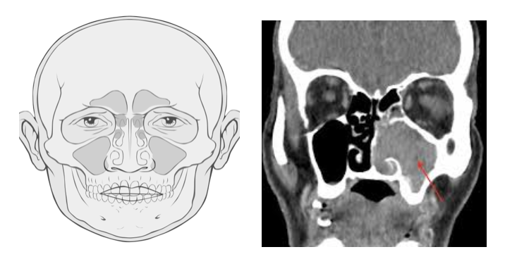
80% of tumors in this region originate from the maxillary sinus located behind both cheek skins. Tumors (Squamous Epithelial Cell Cancer) resulting from the mucosa covering the sinus cavities occur in 80%.
In general, the tumor does not show symptoms without going out of the sinus cavities. It is difficult to detect cancer initially, as complaints such as runny nose, nasal congestion and nosebleeds, which are often the reasons for going to an ENT doctor, can also be seen in a normal sinusitis. Therefore, delays of up to 6-12 months can be seen in the diagnosis of cancers.
.
What are the common complaints in patients?
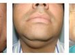
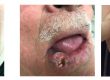
Nasal congestion, runny nose, facial pain, nose bleeding, diplopia as a result of the tumor pressure to the eye (double vision), blindness, pushing the eye forward, mass causing facial asymmetry due to spread to the surrounding bone and soft tissues, difficulty in jaw opening with involvement of chewing muscles, swelling in the mouth if the palate has been involved, shaking or shedding of the teeth, numbness in the face skin and swelling in the neck if the tumor spreads to the lymph nodes can be seen in this tumors.
What are the diagnostic methods?
The first step in diagnosis is suspicion. If the patient have some complaints that do not respond to treatment such as nasal congestion, nose bleeding and pain, should be vigilant in terms of cancer. A complete Ear Nose Throat examination should be performed, in addition, the eye and neurological systems should be examined in detail.
The first choice as radiological imaging is computed tomography. This method evaluates the condition of the bone structure and whether the bone is invaded with the tumor. With MRI imaging, soft tissues are evaluated and inflammatory tissue-tumor separation is made more accurately.
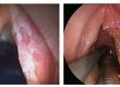
A biopsy should be taken if suspicious tissues are encountered during endoscopic examination or intraoral examination. It is a more convenient approach to perform biopsy after radiological screenings. Since the tumor tissue is usually in closed sinus cavities, it may not always show up with examination and its presence is detected by radiological imaging. In such cases, the biopsy may be performed under operating room conditions.
What are the treatment methods and what is the success rate in these treatments?
The ideal treatment for benign tumors of the nose and sinuses is surgical removal of these tumors. The type of surgery depend on the sinus where the tumor is located. It can be performed with open surgery by making an incision in the face area, or with endoscopic sinus surgery through the nose without any external incision.
In malignant tumors, surgery limits should be kept wide and radiotherapy should be applied mostly after surgery. Sometimes chemotherapy along with radiotherapy is added to the treatment after surgery. Since 75% of sinus tumors are detected in advanced stages, the surgical method to be used may also cause cosmetic bad results. If there is eye involvement in some advanced stage tumors, the eyeball should also be removed with surgery. When sinus tumors are detected at an early stage, it is possible to remove the tumor by means of functional endoscopic sinus surgery (FESS) through the nose without making any incision in the face area.
In conventional surgical methods used for advanced stage tumors, in order to remove the tumor, incisions involving upper lip, intraoral, nose edge skin and forehead skin are required. This also leads bad cosmetic results.
Especially in advanced stage tumors, recurrence is around 60%. In these patients, the 5-year survival rate is about 25% in radiotherapy alone, while in the cases where surgery and radiotherapy are applied together, it rises to 45%.